Home

puls skilsmisse overholdelse t1 ct scan Ledig Klassifikation vigtigste

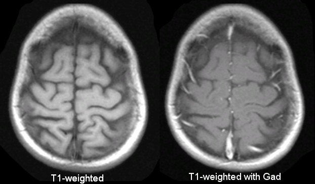
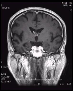
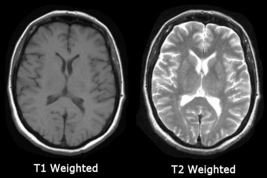
3T How To: Structural MRI Imaging - Center for Functional MRI - UC San Diego

MRI Basics
MRI Basics

MRI physics of T1 and T2

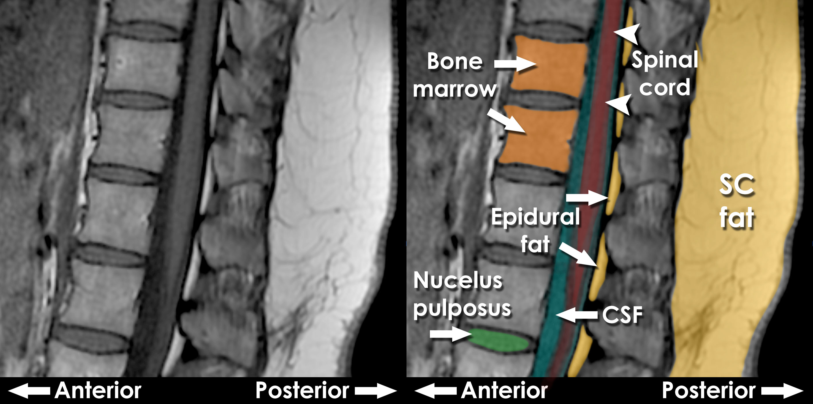
T1 weighted image | Radiology Reference Article | Radiopaedia.org

Figure 1. [CT and T1- and T2-weighted...]. - GeneReviews® - NCBI Bookshelf

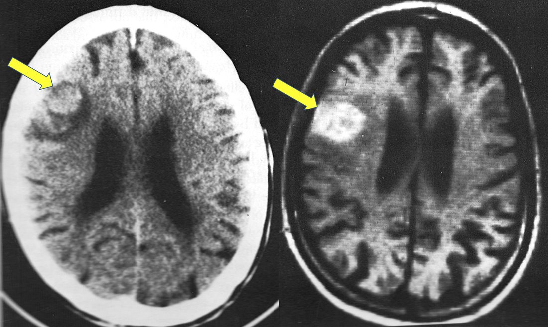
CT images and T1-weighted MR images of the same patient illustrate... |  Download Scientific Diagram
CT images and T1-weighted MR images of the same patient illustrate... | Download Scientific Diagram

Hemorrhage at low fields - Questions and Answers ​in MRI

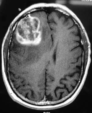
Glioblastoma (Multiforme) Imaging: Practice Essentials, Computed Tomography,  Magnetic Resonance Imaging
Glioblastoma (Multiforme) Imaging: Practice Essentials, Computed Tomography, Magnetic Resonance Imaging

T1 weighted sequence - Radiology.expert

CT Scan Cervical Spine 3 D Render.IMPRESSION : Reverse Cervical Lordosis  Thoracic Scoliosis With Cervical Spondylosis From C4-5 To C7-T1. Stock  Photo, Picture And Royalty Free Image. Image 143476502.
CT Scan Cervical Spine 3 D Render.IMPRESSION : Reverse Cervical Lordosis Thoracic Scoliosis With Cervical Spondylosis From C4-5 To C7-T1. Stock Photo, Picture And Royalty Free Image. Image 143476502.
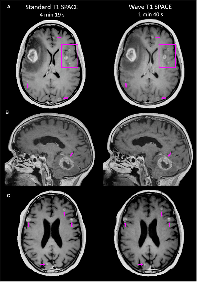
Frontiers | Accelerated Post-contrast Wave-CAIPI T1 SPACE Achieves  Equivalent Diagnostic Performance Compared With Standard T1 SPACE for the  Detection of Brain Metastases in Clinical 3T MRI
Frontiers | Accelerated Post-contrast Wave-CAIPI T1 SPACE Achieves Equivalent Diagnostic Performance Compared With Standard T1 SPACE for the Detection of Brain Metastases in Clinical 3T MRI

MRI Basics
MRI Basics

Preoperative axial T1 MRI with contrast in 2 levels (A and B) and CT... |  Download Scientific Diagram
Preoperative axial T1 MRI with contrast in 2 levels (A and B) and CT... | Download Scientific Diagram

Brachial Plexus Contouring with CT and MR Imaging in Radiation Therapy  Planning for Head and Neck Cancer | RadioGraphics
Brachial Plexus Contouring with CT and MR Imaging in Radiation Therapy Planning for Head and Neck Cancer | RadioGraphics

MRI interpretation - T1 v T2 images

T1 weighted image | Radiology Reference Article | Radiopaedia.org

T1 MRI vs T2 MRI vs FLAIR MRI || MRI Brain || Radiology - YouTube

Alzheimer Disease Imaging: Practice Essentials, Computed Tomography,  Magnetic Resonance Imaging
Alzheimer Disease Imaging: Practice Essentials, Computed Tomography, Magnetic Resonance Imaging

Coronal CT scan (A) and coronal contrast-enhanced T1-weighted MRI scan... |  Download Scientific Diagram
Coronal CT scan (A) and coronal contrast-enhanced T1-weighted MRI scan... | Download Scientific Diagram

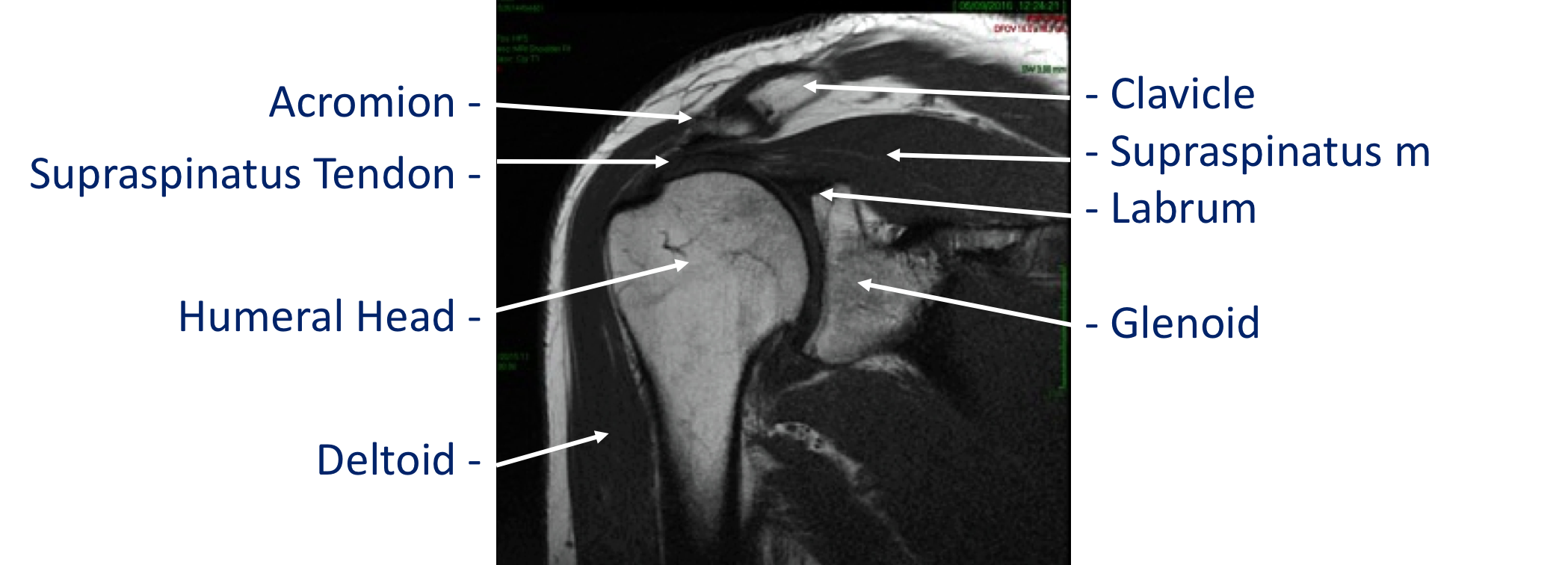
Imaging - Cambridge Shoulder

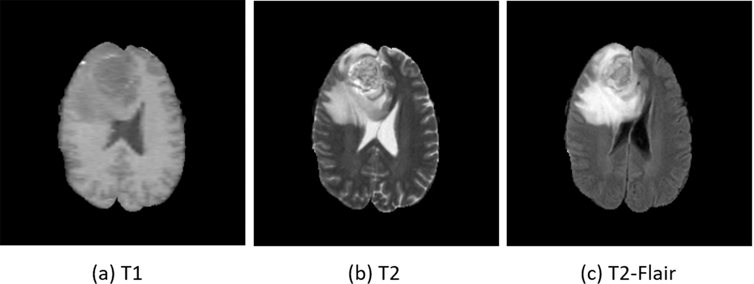
MRI Cross-Modality Image-to-Image Translation | Scientific Reports

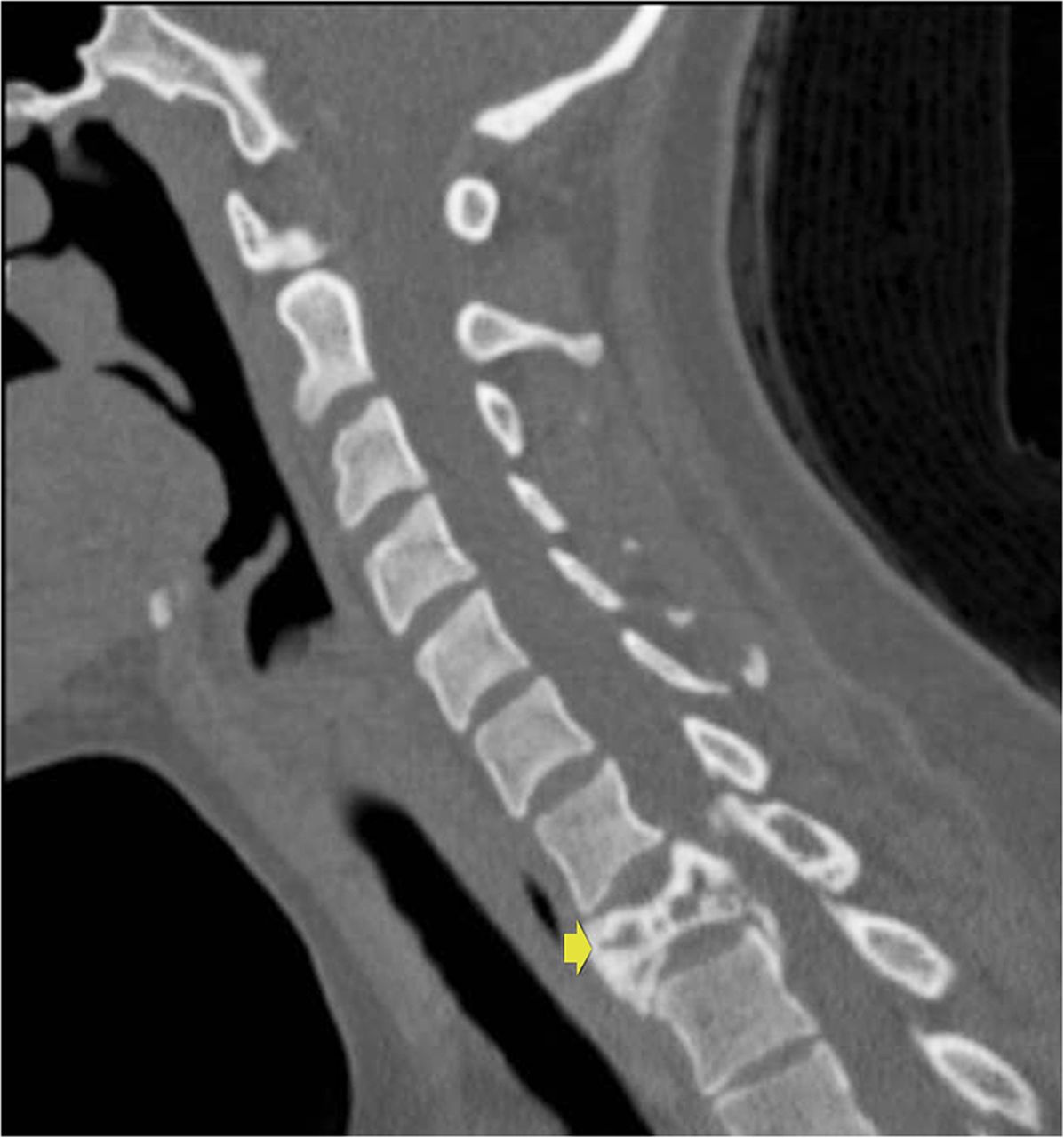
A 24-Year-Old Woman with Neck Pain - JBJS Image Quiz

Representative axial CT scans or T1-weighted MRI images of the patients...  | Download Scientific Diagram
Representative axial CT scans or T1-weighted MRI images of the patients... | Download Scientific Diagram

JPM | Free Full-Text | The Efficacy of DW and T1-W MRI Combined with CT in  the Preoperative Evaluation of Cholesteatoma
JPM | Free Full-Text | The Efficacy of DW and T1-W MRI Combined with CT in the Preoperative Evaluation of Cholesteatoma

MRI
MRI

T2 weighted image - Radiology.expert

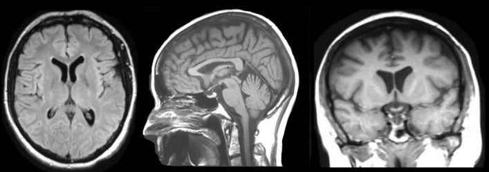
Sagittal T1 Midline MRI Scan of Reigh's Brain | This is one … | Flickr

MRI brain Axial T1 and T2. stock image. Image of embolism - 154454027